Muscles of the Upper Limb
Did you know that each upper limb of the human body has 32 bones and over 40 muscles? The function of these muscles is to create fluid and coordinated movement at the joints of the arm, to assist the ligaments with joint stability, and to provide support and protection for vulnerable arm structures such as nerves, lymphatic and blood vessels.
What are the muscles of the upper limb? The upper limb, also called the upper extremity, is the anatomical region of the body from the clavicle to the distal phalanges of the hands. The muscles of the upper limb originate on the bones of the shoulder girdle and arm. This includes the scapula, clavicle, humerus, radius, ulna, carpal bones, and metacarpals. The muscles of the upper limb create movement at the joints of the shoulder, elbow, wrist, hand and fingers.
This review of the muscles of the upper limb was created to serve as a practical anatomy and kinesiology reference for massage therapists and students preparing to take the FSMTB Massage & Bodywork Licensing Exam (MBLEx). All of the muscles of the upper extremity as well as their origins, insertions, actions and innervations are listed in the two muscle tables below. We’ll also look at muscle groups of the upper extremity, and the similarities and differences between the structure of the upper and lower limbs.


Upper extremity muscles for massage therapists
Muscles are one of the primary types of soft tissues that massage therapists focus on. Manual therapy techniques such as myofascial release, deep tissue massage, neuromuscular massage (NMT), assisted stretching, and Swedish massage are all designed to create positive effects in the muscles and the connective tissues that surrounds them. These benefits include relieving trigger points, decreasing muscle hypertonicity, and reducing adhesions.
As a massage therapist, you’re expected to have a working knowledge of the origins, insertions and actions (OIA) of all major skeletal muscles of the body. And depending on what type of bodywork you provide, it is also helpful to know the fiber directions of these muscles. Since this is information that a massage therapist needs to know to work as a LMT, you can expect to see several questions about muscles on the MBLEx. Test questions on muscle OIA are primarily covered in the kinesiology content area of the massage exam.
***MBLEx study tip: Don’t just study individual muscles. Learn the relative positions of the adjacent muscles and other structures of the surrounding area. Check out some additional study tips for the MBLEx.
You may also be asked basic questions about the muscle innervation on the massage exam. So it is a good idea to be familiar with the large nerves of the upper limb, and know which muscle groups they innervate. For example, have a basic understanding of the function and path of the median nerve, radial nerve, musculocutaneous nerve, axillary nerve, and ulnar nerve. It is unlikely that you will see any questions asking which specific spinal nerves innervate a muscle (e.g. pronator teres is innervated by nerves from C6,7).
Therapists must also be familiar with the endangerment sites for massage. These are areas of the body where certain structures are vulnerable to injury from massage or bodywork techniques. Endangerment sites of the upper limb include the axillary region (also called the axilla or arm pit), the area around the clavicle, the medial side of the upper arm, the anterior part of the elbow joint (antecubital fossa), and the anterior wrist. It is your knowledge of muscle locations and surface anatomy that will help you identify these endangerment sites.
Having a good knowledge of the forearms and hands is particularly important for massage therapists and bodyworkers, since working as a manual therapist can put a lot of strain on these muscles. It is important to always use good body mechanics and avoid putting excessive strain on the small muscles, joints and ligaments of the hands. Therapists should also learn self-care strategies to keep your arms and hands healthy and strong.

Learn Muscles in 60 Days
The Learn Muscles in 60 Days online course is designed to help massage therapy students learn and remember the muscles, origins, insertions and actions for school and for the MBLEx.
Similarities between the upper extremity and lower extremity
A brief comparison of the structure of the upper limbs of the body to the lower limbs reveals similarities between the two regions of the body. The main difference is that the joint structure of the upper extremities is designed for greater mobility, range of motion and motor control. Also, the muscles and bones of the upper limbs are not as large or strong because they do not need to bear the weight of the body like the lower limbs do.
Understanding these similarities will help massage students learning about the structure and function of the musculoskeletal system. Let’s look at some of the similarities between muscles, bones and joints of the upper and lower extremities:
Muscles and bones of the upper limbs
- The upper limb is connected to the axial skeleton by the shoulder girdle
- The joint between the shoulder girdle and the large proximal arm bone (humerus) is a shallow ball-and-socket joint (glenohumeral joint).
- Muscles around this joint create flexion, extension, rotation, abduction and adduction
- There is a hinge joint at the distal end of the humerus (elbow or humeroulnar joint) that permits flexion and extension, which is created by the muscles on the anterior and posterior of the limb
- Just distal to this is the forearm, which is a 2-bone segment that can medially and laterally rotate (pronation & supination)
- These two bones articulate with the carpals bones at their distal end to form the wrist (radiocarpal joint)
- Distal to the wrist is the hand, which contains 27 bones (8 carpals, 5 metacarpals, and 14 phalanges)
- Extrinsic hand muscles and layers of intrinsic muscles create movement of the hand and fingers.
Muscles and bones of the lower limbs
- The lower limb is connected to the axial skeleton by the pelvic girdle
- The joint between the pelvic girdle and the large proximal thigh bone (femur) is a deep ball-and-socket joint (acetabulofemoral joint).
- Muscles around this joint create flexion, extension, rotation, abduction and adduction
- There is a hinge joint at the distal end of the femur (knee or tibiofemoral joint) that permits flexion and extension, which is created by the muscles on the anterior and posterior of the limb
- Just distal to this is the lower leg, which is a 2-bone segment that can medially and laterally rotate
- These two bones articulate with the tarsal bones at their distal end to form the ankle (talocrural joint)
- Distal to the ankle is the foot, which contains 26 bones (7 tarsals, 5 metatarsals, and 14 phalanges) (not counting sesamoid bones)
- Extrinsic foot muscles and layers of intrinsic muscles create movement of the foot and toes.
Overview of the upper extremity muscles and muscle groups
There are several major muscle groups in the upper limb. Recall that muscles can be grouped in different ways. The main way of grouping muscles is based on either their location or their function. Skeletal muscles could also be grouped based on their innervation (e.g. muscles innervated by the radial nerve). Or they could be grouped by the spinal nerve root that innervates them, which is called a myotome (e.g. the C5 myotome is associated with shoulder abduction).
The muscles of the upper limbs are primarily innervated by the nerves of the brachial plexus. These large nerves are mixed nerves, which means that they carry both efferent (motor) nerve signals and afferent (sensory) nerve signals. The nerves of the brachial plexus originate from the spinal cord in the cervical region, specifically C5-T1.
The main function of the arm is to put the hand where it needs to be.
Since full range of motion (ROM) of the upper limb requires paired movement with the scapula, the muscles that control scapular position (periscapular muscles) are involved with all directions of arm movement. For example, full abduction of the arm (reaching overhead) is 180 degrees of motion. The final 60 degrees of this motion comes from upward rotation of the scapula. The muscles that move the scapula, clavicle and humerus which originate on the torso (e.g. periscapular muscles, pectorals, latissimus, serratus anterior) are covered in our Muscles of the Torso review.
Muscle groups of the scapula and shoulder:
- Rotator cuff. Supraspinatus, infraspinatus, teres minor, subscapularis.
- Shoulder flexors. Anterior deltoid, coracobrachialis, biceps brachii (short head), pectoralis major (clavicular head).
- Shoulder extensors. Teres major and minor, posterior deltoid, infraspinatus, triceps (long head). The pectoralis major and latissimus dorsi (muscles of the torso) are also powerful muscles that extend the shoulder (glenohumeral) joint.
- Shoulder abductors. Supraspinatus (initiates GH abduction), and middle deltoid. Full abduction of the arm also involves upward rotation of the scapula, which involves many additional muscles (primarily torso muscles).
Muscle groups of the elbow:
- Elbow flexors. Biceps brachii, brachialis, brachioradialis, pronator teres.
- Elbow extensors. Triceps brachii, anconeus.
Muscle groups of the forearm:
- Forearm pronators. Pronator teres and pronator quadratus.
- Forearm supinators. Supinator, biceps brachii.
Muscle groups of the wrist and hand:
- Wrist flexors. Flexor carpi radialis, flexor carpi ulnaris, palmaris longus.
- Wrist extensors. Extensor carpi radialis longus and brevis, extensor carpi ulnaris.
- Wrist adductors (ulnar deviation). Extensor carpi ulnaris, flexor carpi ulnaris.
- Wrist abductors (radial deviation). Flexor carpi radialis, extensor carpi radialis longus.
- Thenar muscles. Abductor pollicis brevis, flexor pollicis brevis, opponens pollicis.
- Hypothenar muscles. Opponens digiti minimi, abductor digiti minimi, flexor digiti minimi brevis
- Intrinsic muscles of the hand are located entirely within the hand (origin and insertion). These muscles perform the fine motor functions of the hand.
- Extrinsic muscles of the wrist and hand originate in the forearm, and produce gross movements of the wrist and fingers, as well as grip strength.

Complete MBLEx Test Prep!
Enroll in our MBLEx Prep Course for a complete review of muscles, kinesiology, anatomy and all of the topics you need to know to pass the FSMTB massage licensing exam. This organized and convenient course covers all 7 content areas of the massage exam. It is a great resource for current students, or for therapists who have been out of school for years and need a refresher.
Muscles of the Shoulder and Arm
| Muscle | Origin | Insertion | Action | Innervation |
|---|---|---|---|---|
| Anconeus | Posterior side of lateral epicondyle of the humerus | Lateral aspect of olecranon of the ulna | Elbow extension | Radial nerve (C7,8) |
| Biceps brachii | Short head: tip of coracoid process of the scapula Long head: supraglenoid tubercle of the scapula | Tuberosity of the radius, deep fascia of forearm | Both heads flex the elbow and supinate the forearm. Short head also assists with stabilization and flexion of the shoulder joint. | Musculocutaneous nerve (C5,6) |
| Brachialis | Anterior surface of distal half of the humerus | Coronoid process of the ulna, and ulnar tuberosity | Elbow flexion | Musculocutaneous nerve (C5,6) |
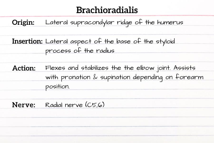
| Brachioradialis | Lateral supracondylar ridge of the humerus | Lateral aspect of the base of the styloid process of the radius | Flexes and stabilizes the elbow joint. Assists with pronation & supination depending on forearm position. | Radial nerve (C5,6) |
| Coracobrachialis | Coracoid process of the scapula | Anteromedial surface of the humerus at mid-shaft | Flexion and adduction of the shoulder joint. | Musculocutaneous nerve (C5-7) |
| Deltoid | Anterior: lateral third of clavicle Middle: lateral acromion Posterior: spine of the scapula | Deltoid tuberosity of the humerus | Anterior: flexion, horizontal adduction and medial (internal) rotation of the humerus at the shoulder joint Middle: abduction of the humerus to 90 degrees Posterior: extension, horizontal abduction and lateral (external) rotation of the humerus | Axillary nerve (C5,6) |
| Infraspinatus | Infraspinous fossa of the scapula | Greater tubercle of the humerus | Lateral rotation of the humerus. Stabilizes the shoulder joint. | Suprascapular nerve (C5,6) |
| Pronator quadratus | Anteromedial surface of the distal ulna | Anterolateral surface of the distal radius | Pronation of the forearm | Median nerve (C8, T1) |
| Pronator teres | Humeral head: medial epicondyle and supracondylar ridge of the humerus Ulnar head: coronoid process of the ulna | Lateral surface of shaft of radius (middle 1/3) | Pronation of the forearm. Elbow flexion. | Anterior interosseous nerve (branch of median nerve) (C6,7) |
| Subscapularis | Subscapular fossa of the scapula | Lesser tubercle of the humerus | Medial (internal) rotation of the humerus. Stabilizes the shoulder joint. | Upper and lower subscapular nerves (C5,6) |
| Supinator | Lateral epicondyle of the humerus, supinator crest of the ulna, radial collateral ligament, annular ligament | Lateral surface of proximal 1/3 of radius, between the anterior and posterior oblique lines | Supination of the forearm at the proximal radioulnar joint. | Deep branch of the radial nerve (C6-8) |
| Supraspinatus | Supraspinous fossa of the scapula | Greater tubercle of the humerus (superior facet) | Initiates abduction of the humerus at the shoulder joint. Stabilization of the head of the humerus in the glenohumeral joint. | Suprascapular nerve (C5,6) |
| Teres major | Inferior angle of the scapula (dorsal surface) | Medial lip of bicipital (intertubercular) groove of humerus | Extension, medial (internal) rotation, and adduction of humerus. | Lower subscapular nerve (C5,6) |
| Teres minor | Upper 2/3 of lateral (axillary) border of posterior surface scapula | Greater tubercle of humerus (inferior facet) | Lateral (external) rotation of humerus. Stabilizes the shoulder joint in the glenoid cavity. | Axillary nerve (C5,6) |
| Triceps brachii | Long head: infraglenoid tubercle of the scapula Lateral head: posterior humerus, proximal to radial groove Medial head: posterior humerus, distal to radial groove | Olecranon process of the ulna | All parts: Extension of the elbow. Long head also adducts and extends the humerus at the shoulder joint. | Radial nerve (C7,8) |

Muscle Flashcards
Can you name the muscle in the image? How about the origin, insertion, and action?
Flashcards are a great way to memorize muscles, anatomy, or other topics while in massage school or when studying for the MBLEx. You can try some of our free MBLEx flashcards. Or you can make flashcards that cover the specific subjects that you need.
Muscles of the Forearm, Wrist and Hand
| Muscle | Origin | Insertion | Action | Innervation |
|---|---|---|---|---|
| Abductor digiti minimi | Pisiform | Base of proximal phalanx of the 5th digit on ulnar side | Abduction and flexion 5th digit (little finger) at MCP joint | Deep branch of the ulnar nerve (C8,T1) |
| Abductor pollicis brevis | Flexor retinaculum, scaphoid and trapezium | Base of the proximal phalanx of thumb (radial side) | Abducts thumb at carpometacarpal joint | Recurrent branch of median nerve (C8,T1) |
| Abductor pollicis longus | Middle third of posterior surface of radius, ulna and interosseous membrane | Base of the first metacarpal (radial side) | Abducts the thumb at carpometacarpal (CMC) joint | Posterior interosseous nerve (C6,7) |
| Adductor pollicis | Oblique head: base of 2nd and 3rd metacarpals (palmar side), capitate Transverse head: shaft of the 3rd metacarpal | Base of proximal phalanx of thumb (medial side) | Adducts the thumb | Deep branch of ulnar nerve (C8, T1) |
| Dorsal interossei of the hand | Each of the 4 muscles arises from the two adjacent metacarpal shafts 1-5 (dorsal side) | Base of the proximal phalanx and the extensor expansion of 2nd, 3rd and 4th digit | Flexion of metacarpophalangeal joint. Extension of DIP & PIP joints of digits 2-4. Abduction of digits 2-4. | Deep branch of ulnar nerve (C8,T1) |
| Extensor carpi radialis brevis (ECRB) | Lateral epicondyle of the humerus via common extensor tendon | Base of the 3rd metacarpal (posterior side) | Extension and abduction (radial deviation) of the wrist and hand. | Radial nerve (C6,7) |
| Extensor carpi radialis longus (ECRL) | Lateral supracondylar ridge of the humerus | Base of 2nd metacarpal bone (posterior side) | Extension and abduction (radial deviation) of the wrist and hand. | Radial nerve (C6,7) |
| Extensor carpi ulnaris | Lateral epicondyle of humerus via common extensor tendon, and posterior border of ulna | Base of 5th metacarpal bone (medial side) | Extension and adduction (ulnar deviation) of the wrist and hand. | Posterior interosseous nerve (C7,8) |
| Extensor digiti minimi | Lateral epicondyle of humerus via common extensor tendon | Merges with extensor digitorum tendon and inserts into the extensor expansion of 5th digit | Extension of metacarpophalangeal (MCP), proximal interphalangeal (PIP) and distal interphalangeal (DIP) joints of the 5th digit | Posterior interosseous nerve (C7,8) |
| Extensor digitorum | Lateral epicondyle of humerus via common extensor tendon | Base of middle phalanges of digits 2-5 (dorsal surface) and extensor expansion of 4 fingers. | Extension of wrist and metacarpophalangeal (MCP), proximal interphalangeal (PIP) and distal interphalangeal (DIP) joints of the 2nd-5th digits. | Posterior interosseous nerve (C7,8) |
| Extensor indicis | Posterior surface of distal ulna and interosseous membrane | Tendon merges with tendon of the extensor digitorum muscle to insert into extensor expansion of index finger (2nd digit). | Extends the index finger at MCP, PIP and DIP joints. Assists with wrist extension. | Posterior interosseous nerve (C7,8) |
| Extensor pollicis brevis | Posterior surface of distal radius and interosseous membrane | Base of the proximal phalanx of the thumb (posterior aspect) | Extension of thumb at MCP joint | Posterior interosseous nerve (C7,8) |
| Extensor pollicis longus | Posterior surface of ulna (middle third) and interosseous membrane | Base of the distal phalanx of the thumb (posterior aspect) | Extension of thumb at metacarpophalangeal (MCP) and interphalangeal (IP) joint | Posterior interosseous nerve (C7,8) |
| Flexor carpi radialis | Medial epicondyle of the humerus via common flexor tendon | Base of the 2nd and 3rd metacarpals | Flexion and abduction (radial deviation) of wrist | Median nerve (C6,7) |
| Flexor carpi ulnaris | Humeral head: medial epicondyle of the humerus via common flexor tendon Ulnar head: olecranon and posterior border of ulna | Pisiform, hook of hamate, and base of 5th metacarpal | Flexion and adduction (ulnar deviation) of wrist | Ulnar nerve (C7-T1) |
| Flexor digiti minimi brevis (hand) | Hook of hamate and flexor retinaculum | Proximal phalanx of 5th digit | Flexion of joints of the 5th digit | Deep branch of ulnar nerve (C8,T1) |
| Flexor digitorum profundus | Anterior and medial surfaces of shaft of ulna (proximal 2/3), interosseous membrane | Base of distal phalanges 2-5 (palmar aspect) | Flexion the MCP, PIP and DIP joints of digits 2-5 | Median nerve (anterior interosseous nerve) (C8,T1) innervates digits 2-3 Ulnar nerve (C8,T1) innervates digits 4-5 |
| Flexor digitorum superficialis | Humeral head: medial epicondyle of humerus via common flexor tendon, and the coronoid process of ulna Radial head: proximal half of anterior border of the radius | Shafts of middle phalanges of digits 2-5 (lateral aspects) | Flexion of metacarpophalangeal (MCP) and proximal interphalangeal (PIP) joints | Median nerve (C8,T1) |
| Flexor pollicis brevis | Superficial head: flexor retinaculum, trapezium Deep head: trapezoid and capitate | Base of proximal phalanx of thumb | Flexion of carpometacarpal (CMC) and metacarpophalangeal (MCP) joints of thumb | Superficial head: median nerve (C6,7) Deep head: ulnar nerve (C8,T1) |
| Flexor pollicis longus | Anterior surface of radius, interosseous membrane | Base of distal phalanx of thumb (palmar surface) | Flexion of the metacarpophalangeal and interphalangeal joints of the thumb | Median nerve (C7,8) |
| Palmar interossei (3) | Shaft of 2nd, 4th and 5th metacarpals (palmar surface) | Base of the proximal phalanges and extensor expansion of 2nd, 4th and 5th digits | Flexion and adduction of 2nd, 4th and 5th MCP joints | Deep branch of ulnar nerve (C8,T1) |
| Lumbricals of the hand (4) | Flexor digitorum profundus tendons of digits 2-5 | Extensor expansion of digits 2-5 at proximal phalanges (radial side) | Flexion of metacarpophalangeal joints, and extension of proximal and distal IP joints of digits 2-5 | Lumbricals 1-2: median nerve (C6,7) Lumbricals 3-4: ulnar nerve (C8) |
| Opponens digiti minimi | Hook of hamate and flexor retinaculum | Shaft of 5th metacarpal (ulnar side) | Opposition of 5th digit at CMC joint | Deep branch of ulnar nerve (C8,T1) |
| Opponens pollicis | Trapezium and flexor retinaculum | Shaft of 1st metacarpal (radial side) | Opposition of thumb at CMC joint | Recurrent branch of median nerve (C6,7) |
| Palmaris brevis | Palmar aponeurosis | Dermis of skin of palm at hypothenar region | Draws the skin of ulnar side of hand toward the center of the palm. Tightens palmar aponeurosis and grip | Superficial branch of ulnar nerve (C8,T1) |
| Palmaris longus | Medial epicondyle of the humerus via common flexor tendon | Palmar aponeurosis | Flexion of wrist | Median nerve (C7,8) |

Join MBLEx Guide Today!
Become a member of MBLExGuide to get access to the best online MBLEx test prep resources on the market. Our full-length practice exams, rapid quizzes and complete MBLEx Prep Course will help you pass the massage exam with confidence and begin working as an LMT.