Annalie Bamberg
Spring 2024. East West College of the Healing Arts. Hillsboro, Oregon
The closer I come to earning the title of LMT, the more I look forward to building a career around supporting others in their healing by addressing what is stored somatically. I’ve seen massage work as an amazing modality for healing. As a young child, I watched my mama go through massage school and build her practice, and starting in my teens I experienced somatic healing for myself. I’m excited to get to support others in their healing journeys as well. The biomechanical aspect of massage adds a layer that, as a dancer, I find intriguing and beautiful. My massage career will incorporate my background as a dancer with my massage school education to create a versatile approach to client care. There is so much to learn, and I look forward to gaining experience and furthering my knowledge. I hope to contribute to research in the future, so this case study application was a good start for me! I plan to start out doing therapeutic massage in a clinical or wellness setting while working toward becoming a Doctor of acupuncture and Chinese medicine, which will increase the tools I have available in providing client care. I’m grateful to receive these scholarship funds, which will help me to pay for tuition in my final term this fall.
Case Study: Myofascial release along fascial trains may reduce forearm tension related to fine motor function
Abstract
This study provides evidence in favor of the use of myofascial release in decreasing forearm tension, and suggests that fascial lines may provide a guide for effective treatment areas. Myofascial release is a massage modality that focuses on releasing restrictions within the fascial sheets of the musculoskeletal system. These fascial sheets are present within the entire musculoskeletal system, including the forearms. Research indicates that restriction within this fascial system, both locally and distant to observable symptoms, may be related to its role in motor activities (Garofoli and Svanera; Myers, Thomas). A client presenting with forearm tension and restricted wrist movement reports an increase in symptoms with performance of fine motor activities. The client received myofascial release treatment along the fascial arm lines as they are presented in Anatomy Trains (Myers, Thomas). At the end of the treatment plan, tension reduction was seen in NRS-11 pain scale ratings as reported by the client and movement was increased based on therapist assessment of PROM. Based on these results, myofascial release may be an effective treatment for forearm tension and restricted wrist movement. Further research is indicated.
Introduction
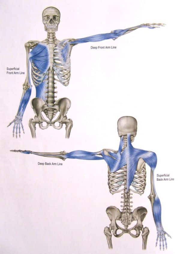
Until fairly recently, studies into movement and motor function have focused largely on the role of muscle tissue. Recent evidence, such as a 2019 study published by the European Journal of Translational Myology (Garofoli and Svanera), suggests that fascia and other connective tissues may play an equally critical role when it comes to motor control. The patterns of this role are explored in depth in the book Anatomy Trains (Myers, Thomas). Anatomy Trains is based on research findings that indicate fascia, the connective tissue surrounding the musculoskeletal system, to be a continuous sheet within the body. The book outlines the resulting relationships in detail. For the purposes of this study, we will be focusing on the fascial relationships within the arms (figure 1).
These relationships mean that restriction within an area of the fascia may cause pain or dysfunction in a distant area of the body, because the areas are connected through fascial lines. In the case of the fascial arm lines, we see that the fascial sheets of the forearms are connected all the way up to the sternum and spine. There are four arm lines; the superficial front (SFAL), deep front (DFAL), superficial back (SBAL), and deep back (DBAL). The SFAL includes pectoralis major, latissimus dorsi, teres major, medial intermuscular septum, forearm flexor group, and anterior phalangeal tendons. The DFAL includes pec minor, subclavius, biceps brachii, coracobrachialis, periosteum of the radius, radial collateral ligament, and the pollicis muscles. The SBAL includes trapezius, deltoids, lateral intermuscular septum, forearm extensor group, and posterior phalangeal tendons. The DBAL includes rhomboids, infraspinatus, teres minor, rectus capitis, levator scapula, supraspinatus, subscapularis, triceps brachii, periosteum of the the ulna, and the ulnar collateral ligament. The medial and lateral intermuscular septum is a connective tissue structure that separates the flexors and extensors of the arms.
Within the forearms there are upwards of 15 muscles, and the majority of these muscles are divided into the flexor and extensor groups. The flexor group includes the muscles responsible for flexion of the wrist and fingers such as flexor carpi radialis and ulnaris and flexor digitorum superficialis and profundus. The extensor group includes the muscles responsible for extension of the wrist and fingers such as extensor carpi radialis longus and brevis, extensor carpi ulnaris, and extensor digitorum. The forearm muscles responsible for adduction of the wrist are located on the ulnar side, such as extensor carpi ulnaris and flexor carpi unlnaris. Accordingly, the muscles responsible for abduction of the wrist are located on the radial side, such as extensor carpi radialis brevis and longus, and flexor carpi radialis. These muscles of the forearms and their actions of flexion, extension, adduction, and abduction of the wrist and fingers are those required for fine motor activities. Because their fascial sheets are connected all the way up the arms lines, as far as trapezius, pectorals, and latissimus dorsi, fascial restrictions anywhere along the arm lines may impact the function of the forearm muscles. This restriction within the arm lines can be seen in a client as impairment of or pain with fine motor function. From the client’s perspective, this restriction within the fascia can be perceived as tension. The terms restriction and tension will be used interchangeably throughout this study. These concepts of fascial relationships and their role in motor control have important implications for massage therapy, in which a primary focus is the manipulation of soft tissues to address movement and overuse-based tension and injury.
Behind this case study is the question, “What is the impact of myofascial release along fascial lines of the arm in the context of forearm tension related to fine motor function?” Fine motor function includes precise movements that require the use of small, specialized muscles (“Fine Motor Coordination”). In this study, we use the term fine motor function to refer to precise movements of the wrist and hand such as twisting or grasping . This study will focus on addressing forearm tension by using myofascial release along the fascial lines of the arms as they are portrayed in Anatomy Trains. Myofascial release is defined by Art Riggs in Modalities for Massage and Bodywork as “a collection of approaches and techniques that focus on freeing restrictions of movement that originate in the soft tissues of the body” (Stillerman). In this case study, soft tissues will refer specifically to connective tissue sheets around and within the musculoskeletal system. The client’s forearm tension increases with fine motor activities, suggesting movement-based causes. By addressing this complaint with myofascial release along the fascial lines, this study seeks to explore the effectiveness of connective tissue focused treatment, specifically along the fascial lines in response to a movement and overuse-based complaint.

Fig 1. Fascial trains of the arms
Client Details
The client is a 46 year old male presenting with bilateral forearm tension in both the anterior and posterior forearm compartments. The client spends significant amounts of time doing mechanical work as a mechanical technician, with tasks involving fine motor skills performed intermittently throughout a twelve hour shift. Such tasks include repair of microchip machinery and data entry. Machinery repair requires the client to deconstruct and reconstruct small pieces of machinery using their own hands, a screwdriver, and other small tools. Data entry requires keyboard typing. Other daily activities include typing on a personal computer, phone, and gaming system. Previously, he was an automotive technician for 30 years. This position required similar deconstructive and reconstructive activities to microchip machinery repair, done constantly over an eight-hour shift.
The client reports forearm tension increases when both work and daily activities are performed. This tension is usually experienced as tightness throughout the anterior and posterior forearm. Sometimes stiffness in the fingers is experienced as well. Tension begins with any of the activities described above and increases with increased duration of the activity. Tension increases over the 24 hours immediately following cease of the aggravating activities, and this is usually when stiffness in the fingers is experienced. Following this 24-hour period, the tension decreases with rest. The client experiences greater tension in his right arm. He is right-hand dominant. The client denies any health history that indicates underlying health issues or past injuries that may contribute to his current condition.
Assessment
The client’s goal is to decrease forearm tension, which worsens with activity. Assessments focused on tension rating and range of motion (ROM) observations in addition to palpation. To begin, the client rated his tension using the Numeric Pain Rating Scale (NRS-11), with 0 being no tension and 10 being extreme tension. After obtaining an NRS-11 rating, the client’s wrist range of motion was assessed, both active (AROM) and passive (PROM) with a range of within normal limits (WNL), light, moderate, and severe. ROM assessments were performed for flexion, extension, adduction, and abduction of the wrist. Average ROM for the wrist is considered 80 degrees of flexion, 70 degrees of extension, 30 degrees of adduction, and 20 degrees of abduction. NRS-11 rating, AROM and PROM, and palpatory assessments were all performed at the start and end of each session. Both the pain scale rating and the ROM assessments will be considered objective measurements to assess the effectiveness of the interventions.
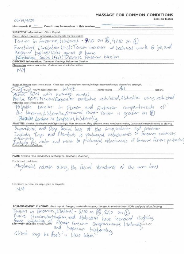
At the start of the first session, the client reported an NRS-11 rating of 7/10 on the right, and 4/10 on the left. AROM assessed as WNL. Passive flexion and extension of wrist assessed as lightly restricted, and passive abduction assessed as moderately restricted. Passive adduction assessed as WNL. Palpation along the fascial arm lines revealed greatest areas of restriction in the proximal third of the flexor and extensor compartments of the forearm, and the trapezius. At the end of the session, the client reported an NRS-11 rating of 5/10 on the right and 2/10 on the left. AROM assessed as WNL. Passive flexion and extension assessed WNL and abduction assessed as moderately restricted. Passive adduction remained WNL. Palpation along the fascial arm lines revealed decreased restriction in the proximal third of the flexor compartment of the forearm and the trapezius, and no change to restriction in the proximal third of the extensor compartment of the forearm.
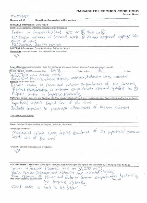
At the start of the second session, the client reported an NRS-11 rating of 5/10 on the right and 3/10 on the left. AROM assessed as WNL. Passive flexion and extension assessed as lightly restricted, passive abduction assessed as moderately restricted. Passive adduction assessed as WNL. Palpation along the fascial arm lines revealed greatest restriction in the proximal third of the extensor compartment of the forearm, followed by areas of restriction in the flexor compartment of the forearm and the trapezius. At the end of the session, the client reported an NRS-11 rating of 5/10 on the right and 2/10 on the left. AROM assessed as WNL. Passive flexion and extension assessed as WNL, and passive abduction assessed as lightly restricted. Passive adduction remained WNL. Palpation along the fascial arm lines revealed decreased restriction in the proximal third of the flexor and extensor compartments of the forearm and the trapezius. Palpable restriction in the extensor compartment remained the greatest.
Treatment Plan
The client will receive two 50-minute sessions one week apart. These sessions will utilize myofascial release techniques along the arm lines (figure 1) as they are represented in Anatomy Trains. Treatment will be focused on releasing restrictions within the fascial sheets of the trapezius, levator scapula, latissimus dorsi, deltoids, pectorals, triceps, biceps, forearm flexors, and forearm extensors. These fascial sheets include the medial and lateral intermuscular septum and all tendinous attachments of the muscles named above. Myofascial techniques may include pin and stretch, cross hand stretch, C-bowing, S-bowing, freeing from entrapment, passive joint mobilization, and arm pulling. It will be suggested that the client increase their water intake, especially for the 24 hours following each session. The client may also incorporate gentle passive stretches of the forearm throughout their day. Passive stretching would include using one hand to hold the second through fifth fingers in extension and passively moving their opposite wrist through flexion, extension, abduction, and adduction. Each stretch should be done for 60 seconds. This may include a 60 second static hold or sets of two, three, or four holds that add up to 60 seconds (Harvard Health Publishing). Stretches were demonstrated for the client. Passive stretching will ensure that the focus is on increasing connective tissue pliability as opposed to muscular tissue. The overall goal of the sessions is to decrease forearm restriction by increasing connective tissue pliability. In the short term, this may look like decreased tension immediately after each session as reported by the client and assessed by the therapist. In the long term, this may look like decreased tension after performance of fine motor activities at work and home, experienced by the client as tightness in the forearms and stiffness in the fingers. For the sake of this case study, only the short term goal will be measured through assessment of tension immediately before and after each session.
Treatment Sessions
Session 1: May 15th 2024, 50 minutes
Myofascial release was applied to all four arm lines (superficial front, deep front, superficial back, deep back) as outlined in Anatomy Trains (figure 1). Ten minutes of broad fascial release including pin and stretch, cross hand stretch, and joint mobilization techniques was performed bilaterally over the trapezius, levator scapula, latissimus dorsi, deltoid, triceps, forearm flexors, and forearm extensors. Five minutes were spent using C-bowing and S-bowing techniques for specific fascial release through bilateral trapezius and latissimus dorsi. Five minutes were spent using C-bowing, S-bowing, freeing from entrapment techniques, cross hand stretch, and pin and stretch techniques on each forearm flexor group. Client was turned to supine at 25 minutes. Five minutes were spent applying pin and stretch and joint mobilization techniques bilaterally to the pectorals, biceps, forearm flexors, and forearm extensors. Eight minutes were spent using C-bowing, S-bowing, freeing from entrapment, pin and stretch, and cross hand stretch techniques on each forearm extensor group. Four minutes were spent on passive flexion, extension, adduction, and abduction of the wrists and passive flexion and extension of the fingers bilaterally. The client reported feeling slight stretching sensations during the treatment, and some involuntary twitching of the fingers occurred. Client reported that overall forearm restriction was decreased from 7/10 to 5/10 on right, and 4/10 to 2/10 on left. The forearm extensor compartments remained the most tense. Passive flexion, extension, and abduction increased slightly, no change to AROM.

Figure 2. Session Notes 05/15/2024
Session 2: May 22nd 2024, 50min
Myofascial release was applied to the superficial back arm line, the line that goes through the extensor compartment of the forearm (figure 1). Twenty minutes were spent on broad fascial release including pin and stretch, cross hand stretch, passive joint mobilization, and arm pulling techniques of bilateral trapezius, latissimus dorsi, deltoids, and forearm extensors. The client was turned to supine at 20 minutes. Twelve minutes were spent using C-bowing, S-bowing, freeing from entrapment, pin and stretch, and cross hand stretch techniques on each forearm extensor group. Six minutes were spent on passive flexion, extension, adduction, and abduction of the wrists and passive flexion and extension of the fingers bilaterally. The client reported feeling slight stretching sensations, and some involuntary twitching of the fingers occurred during treatment. At the beginning of the session, Client reported tension of 5/10 on the right and 3/10 on the left. At the end of the session, client reported tension of 5/10 of the right and 2/10 on the left. The forearm extensor compartments remained the most tense. Passive flexion, extension, and abduction increased slightly, no change to AROM.

Figure 3. Session Notes 05/22/2024
Results and Observations
Upon completion of the treatment plan, the client reported an NRS-11 rating of 5/10 on the right, and 2/10 on the left. This is a difference of two points on both the right and the left, having started at 7/10 and 4/10 respectively. AROM, which appeared average at the start of treatment, showed no change. Passive flexion, extension, and abduction all increased. At the start of the treatment, passive flexion and extension were lightly restricted, and passive abduction was moderately restricted. Upon completion of treatment, passive flexion and extension were WNL, and passive abduction remained lightly restricted. Palpation revealed decreased restriction within the forearm flexors and extensors, and the trapezius. Prior to treatment, this restriction was palpated as tissue that was stiff, resistant to movement, and adhered to nearby structures such as bone or the fascial sheets of other muscles. Post-treatment, the same areas of tissue were found to be softer, with increased pliability and greater movement independent of other structures. The treatment plan was adjusted after the first session based on the finding of greatest tension in the extensor compartments of the forearms. While the first session addressed structures within all four fascial arm lines, the second session was modified to address only structures within the superficial back arm line, which included trapezius and the forearm extensors, in order to give more specific attention to the extensor compartments.
The first session, focusing on myofascial release within all four arm lines, showed the greatest amount of improvement. This session resulted in changes to the client’s reported NRS-11 rating of 7/10 to 5/10 on the right, and 4/10 to 2/10 on the left. This session also showed increases in passive flexion and extension, and abduction. At the conclusion of the first session, the client reported that the tension improvement applied to all areas except the extensor compartment.
The second session, focusing on myofascial release within the superficial back arm line in order to address the extensor compartment restriction more specifically, showed less objectively significant improvement. At the start of the session, the client reported an NRS-11 rating of 5/10 on the right and 3/10 on the left. At the end of the session, the client reported an NRS-11 rating of 5/10 on the right and 3/10 on the left. This session showed increases in passive flexion, extension, and abduction. Passive abduction remained the most restricted.
Upon completion of the treatment plan, the client stated that his forearm tension was “a little better” and that he believed it would improve more if he received additional consistent treatment.
Conclusion
Results of this study indicate that myofascial release may be an effective treatment in reducing movement-based muscular tension in the forearm, and that fascial lines may be a valuable guide in forming an effective treatment plan. The client complained of tension in the forearms that increased with fine motor activities such as typing, and holding or screwing small machine pieces. Myofascial release was performed both locally to the symptoms and on connected structures based on the guidance of Anatomy Trains. Specifically, fascial release was performed on trapezius, levator scapula, latissimus dorsi, deltoids, pectorals, triceps, biceps, forearm flexors, and forearm extensors. Interestingly, the greatest amount of fascial adhesions were found and released within the trapezius and the forearm flexor and extensor compartments. This application of myofascial release along the fascial lines of the arms saw changes in both client reported NRS-11 rating for forearm tension and PROM of the wrist as assessed by the therapist. Additionally, the therapist noted a reduction in palpable restriction within the extensor and flexor compartments of the forearms as well as the trapezius, felt as increased pliability and freedom of movement. These results are in line with the concepts presented by Thomas Myers within Anatomy Trains, and by Garofolini and Svanera with their article on the relationship between fascia and motor function. Significantly, the aspect of trapezius restriction being found and released suggests effective application of the fascial trains theory, as the trapezius is part of the same fascial line (SBAL) as the forearm extensor compartments where the greatest restriction was found. However, it should be noted that the second session, which addressed only the SBAL, achieved less change of client reported NRS-11 rating than the first session, which focused on all four arm lines (SFAL, DFAL, SBAL, DBAL). Future studies should seek to explore this finding further. Overall, results indicate that the concepts of fascial train and fascial and motor function relationships may be effectively applied by manual therapy practitioners who address movement-based tension, however, additional studies should be conducted to verify and expand on results.
The applicability of these concepts is intriguing for several reasons. Massage therapists tend to see many clients with goals of reducing pain with motor function. If fascia is equally important as muscular tissue in its role in motor function, massage therapists may have reason to pay greater attention to the fascia in order to achieve successful rehabilitation. Additionally, every massage therapist is aware that addressing the root cause of the problem is key to obtaining results. This root cause can be challenging to find. By describing structural relationships of the fascia, Anatomy Trains suggests that the root cause may not be located in the same area of the body as the symptoms that it causes, and provides a guide to locating the cause based on the symptom. These concepts of fascial role in motor function and the fascial train relationships could mean additional tools in a massage therapist’s toolbox. Additional tools may also mean increased effectiveness in client treatment, especially because myofascial release has the benefit of being a gentle technique. Some bodies do not have the capacity to handle intense or rigorous bodywork. In these cases, having gentler techniques on hand that are equally effective in meeting client goals is critical to successful client treatment.
This case study is limited by its small size and short duration, having followed only one client through two consecutive sessions over the course of 2 weeks. More expansive studies should be considered to further confirm and expand on the results of this study. Larger numbers of participants and longer tracking times would increase the significance of results. This case study also assessed only short term outcomes by assessing NRS-11 tension rating and PROM immediately before and after each session. Assessment of long term outcomes would provide additional insight into treatment effectiveness.
Future case studies should also consider exploring treatments comparatively. This could include comparing modalities, treatment durations, and areas of treatment application. For example, myofascial release techniques utilized in this case study could be compared to a treatment of only passive stretches, or a treatment utilizing proprioceptive neuromuscular facilitation (PNF) techniques such as contract-relax-antagonist contract (CRAC). How do the impacts of these treatments differ? What does this imply for myofascial release as a treatment option? Treatment plans could also be compared, which could include differing number of sessions, time between sessions, or sessions lengths. What durations of myofascial release application are most effective in achieving client goals? Areas of application can be compared as well, in order to further explore the application of the fascial trains guidance. This could include comparing treatment along the fascial trains with treatment only at the site of symptoms. It could also include comparing treatment along different structures. For example, the treatment plan of this case study could be recreated, with the exception of comparing application only along the SBAL to application along both the SBAL and SFAL. What areas of application for myofascial release are most effective in meeting client goals? These comparative treatment studies should also take into consideration client demographics. Are there any themes? What demographic differences are there in client response to differing modalities, treatment durations, or areas of treatment application? Exploration of such questions surrounding treatment approaches would assist in ascertaining the most effective approaches for different clients, and this knowledge would provide massage therapists with a valuable tool in treating clients more effectively.
Works Cited
“Fine Motor Coordination.” TheFreeDictionary.com, medical-dictionary.thefreedictionary.com/fine+motor+coordination. Accessed 22 June 2024.
Garofolini, Alessandro, and Daris Svanera. “Fascial Organisation of Motor Synergies: A Hypothesis.” European Journal of Translational Myology, vol. 29, no. 3, 8 Aug. 2019, https://doi.org/10.4081/ejtm.2019.8313. Accessed 5 Mar. 2023.
Harvard Health Publishing. “The Ideal Stretching Routine – Harvard Health.” Harvard Health, Harvard Health, 2019, www.health.harvard.edu/staying-healthy/the-ideal-stretching-routine.
Myers, Thomas W. Anatomy Trains : Myofascial Meridians for Manual and Movement Therapists. Edinburgh ; New York, Elsevier, 2009.
Stillerman, Elaine. Modalities for Massage and Bodywork. St. Louis, Missouri, Elsevier/Mosby, 2016.