Muscle Review – Trapezius, Levator scapula, Latissimus Dorsi, Teres major
MBLEx Muscle Review
Introduction
The kinesiology section of each lesson in this MBLEx Prep Course is followed by a Muscle Review. A total of 114 muscles are covered in this course. Each review includes the muscle’s Origin, Insertion, Actions, and Nerve (OINA). Additional notes and study tips are included to help reinforce your understanding, boost retention, and improve your ability to apply this knowledge in clinical settings.
Study Tip:
When reviewing each muscle, try to visualize not only the muscle itself but also the surrounding structures. Consider:
- Which muscles lie deep to it? Which ones are superficial?
- What muscles are medial, lateral, proximal, or distal to the illustrated muscle?
- Is the muscle part of a functional group? (e.g., semitendinosus is part of the hamstrings)
- What are its synergists and antagonists?
- How does it contribute to movement?
A study method that many students find useful is to create your own flashcards using blank index cards. Writing the information in your own words and sketching simple illustrations engages active learning. Doing this as you work your way through the MBLEx Prep Course will significantly improve retention.
While it is unlikely that you will see more than one or two questions on muscle innervation, learning them can still be valuable. Understanding muscle innervations will help when learning dermatomes and myotomes.
👉 Tip: Get familiar with the major peripheral nerves of the body, such as the radial, median, ulnar, axillary, sciatic, femoral, tibial, and fibular nerves.

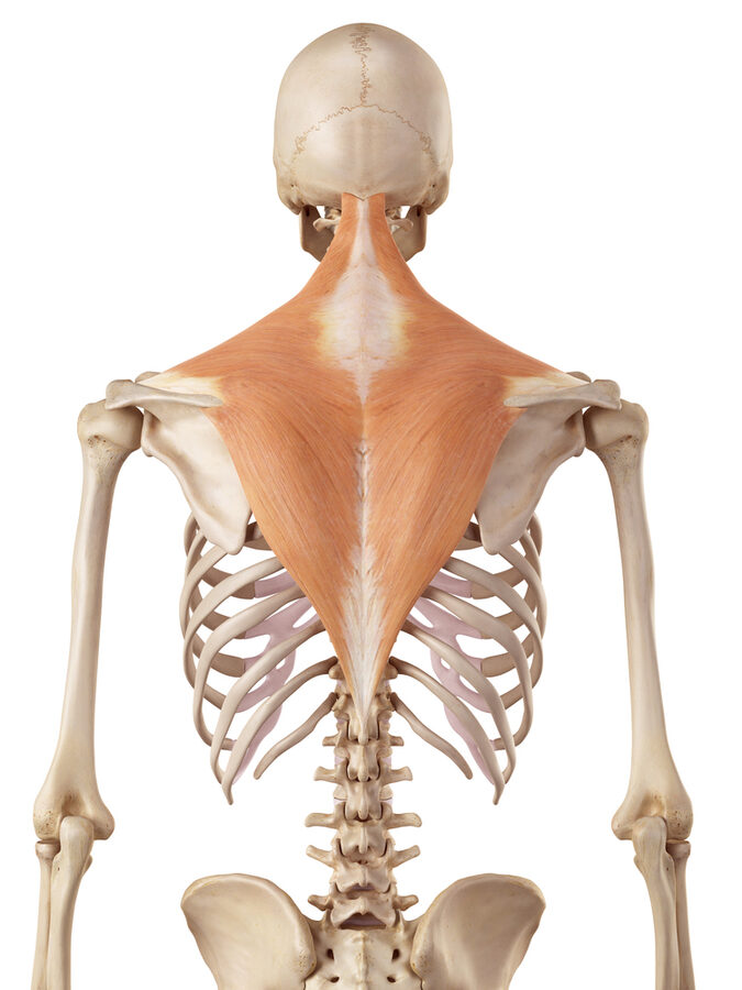


Trapezius
Origin: External occipital protuberance, medial third of the superior nuchal line, ligamentum nuchae, spinous processes of C7-T12
Insertion: Lateral third of the clavicle, acromion process, and upper crest of the scapular spine
Action:
- Upper fibers: Elevate scapula, assist in cervical extension
- Middle fibers: Retract scapula (adduction)
- Lower fibers: Depress and upwardly rotate scapula
Innervation: Accessory nerve (CN XI), cervical nerves (C3, C4)
Notes & Tips
Palpate the upper trapezius by placing your fingers just superior to the clavicle and lateral to the cervical spine, near the posterior aspect of the shoulder. The middle and lower fibers are thinner and harder to isolate. When studying, visualize how fiber direction affects function—upper fibers pull up, middle retract, and lower depress. The trapezius is sometimes confused with the levator scapulae, which also elevates the scapula but does not retract it. Its innervation (CN XI, 11th cranial nerve) is unique compared to most back muscles, which are usually innervated by spinal nerves.


Levator Scapulae
Origin: Transverse processes of C1-C4
Insertion: Medial (vertebral) border of the scapula, between the superior angle and the scapular spine
Action: Elevation and downward rotation of the scapula
Innervation: Dorsal scapular nerve (C5), cervical nerves (C3, C4)
Notes & Tips
The levator scapulae is named after its function—it elevates the scapula. It runs diagonally along the side of the neck, making it easy to palpate near the superior angle of the scapula.
This muscle often becomes tight due to forward-head posture, which can contribute to neck pain and stiffness. Stretching and myofascial release techniques can help alleviate tension.


Latissimus Dorsi
Origin: Spinous processes of T7-L5, sacrum, thoracolumbar fascia, posterior iliac crest, lower 3 or 4 ribs
Insertion: Intertubercular (bicipital) groove of the humerus
Action: Extension, adduction, and medial (internal) rotation of the humerus at the glenohumeral joint
Innervation: Thoracodorsal nerve (C6, C7, C8)
Notes & Tips
The latissimus dorsi is one of the largest muscles in the upper body and plays a key role in pulling movements such as pull-ups, swimming strokes, and rowing motions. It is sometimes referred to as the “swimmer’s muscle” due to its importance in arm extension and adduction. It can be palpated along the posterior and lateral thoracic region, extending toward its insertion on the proximal humerus.
👉 Tip: The latissimus dorsi works synergistically with the teres major to perform similar functions—both extend, adduct, and medially rotate the humerus.


Teres Major
Origin: Inferior angle of the scapula (dorsal surface)
Insertion: Medial lip of the intertubercular (bicipital) groove of the humerus
Action: Extension, medial (internal) rotation, and adduction of the humerus
Innervation: Lower subscapular nerve (C5, C6)
Notes & Tips
The teres major is sometimes called the “lat’s little helper” because it functions almost identically to the latissimus dorsi. It shares an insertion site with the latissimus dorsi and assists in the same motions.
Palpation Tip: The teres major is located just inferior to the teres minor and can be palpated along the posterior axillary border, especially when the arm is actively resisted into internal rotation.