Kinesiology – Concepts & Definitions
Kinesiology Concepts & Definitions

Anatomical Directional Terms
Directional terms help describe the location of structures in relation to other structures. These terms are often paired for clarity and precision. Below are key terms to know for the MBLEx:
Anterior: In front of; toward the front (e.g., the sternum is anterior to the spine).
Posterior: Toward the back or rear (e.g., the scapula is posterior to the ribs).
Superior: Above or over (used for the trunk, head, and pelvis). Example: The nose is superior to the mouth.
Inferior: Below or under (used for the trunk, head, and pelvis). Example: The navel is inferior to the sternum.
Proximal: Closer to the point of origin (used for extremities). Example: The shoulder is proximal to the elbow.
Distal: Farther from the point of origin (used for extremities). Example: The fingers are distal to the wrist.
Superficial: Closer to the surface. Example: The skin is superficial to the muscles.
Deep: Below or farther from the surface. Example: The femur is deep to the quadriceps muscle.
Lateral: Away from the midline; toward the side. Example: The ears are lateral to the nose.
Medial: Toward the midline of the body. Example: The sternum is medial to the shoulders.
Bilateral: On or involving both sides of the body. Example: The kidneys are bilateral structures.
Unilateral: On or involving only one side of the body. Example: A stroke affecting only one hemisphere of the brain can cause unilateral paralysis.
Dorsal: Toward the back. Example: The spine is on the dorsal side of the body.
Ventral: Toward the anterior torso or belly. Example: The navel is on the ventral side of the body.
Radial: Lateral (thumb) side of the forearm or hand. Example: The radial artery is palpable at the wrist near the thumb.
Ulnar: Medial side of the forearm or hand. Example: The ulnar nerve runs along the medial side of the forearm.
Ipsilateral: On the same side of the body. Example: The right hand and right foot are ipsilateral.
Contralateral: On the opposite side of the body. Example: The left brain controls contralateral motor function, meaning it controls the right side of the body.
Some anatomical terms combine two directional references to describe a location more precisely. These terms help pinpoint structures in relation to multiple planes of the body, making anatomical descriptions more specific and useful in clinical communication and documentation. Examples include:
- Anterolateral – Both anterior and lateral
- Anteromedial – Both anterior and medial
- Posterolateral – Both posterior and lateral
- Posteromedial – Both posterior and medial
- Superolateral – Both superior and lateral
- Superomedial – Both superior and medial
- Inferolateral – Both inferior and lateral
- Inferomedial – Both inferior and medial
Other movement terms include:
Deviation: Movement away from the standard position (e.g., ulnar deviation of the wrist).
Tilt: Angling a structure without rotation (e.g., head tilt).
Hyperextension: Extension beyond the normal range of motion (e.g., knee hyperextension).
Circumduction: Circular movement involving multiple joint motions (e.g., arm circumduction at the shoulder).
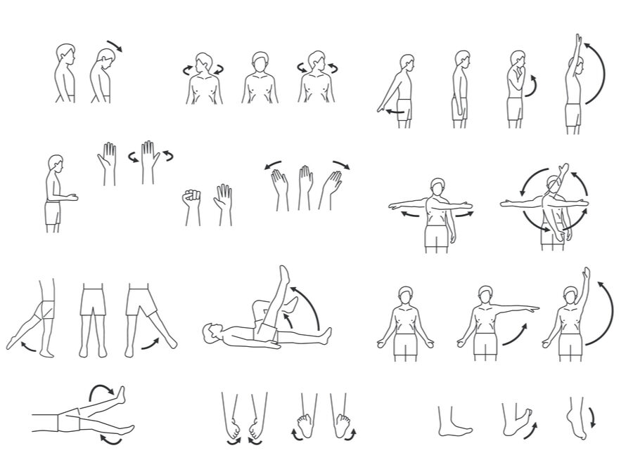
Types of Movement
These are fundamental movements of the body, often grouped in pairs of opposing motions. We’ll explore specific examples in future lessons when discussing joints, movement, and range of motion.
Flexion / Extension: Decreasing or increasing the angle of a joint (e.g., bending and straightening the elbow).
Elevation / Depression: Raising or lowering a structure (e.g., shrugging and lowering the shoulders).
Medial / Lateral Rotation (Also called Internal / External Rotation): Rotating toward or away from the midline (e.g., rotating the humerus inward or outward at the shoulder joint).
Right / Left Lateral Flexion: Bending the trunk or neck sideways (also called side bending).
Abduction / Adduction: Moving a limb away from or toward the midline (e.g., spreading and closing the fingers).
Horizontal Abduction / Adduction: Moving a limb away from or toward the midline in the transverse plane (e.g., moving the arm outward or inward while it is at shoulder height).
Dorsiflexion / Plantarflexion: Flexing or extending the foot at the ankle (e.g., lifting toes upward vs. pointing toes downward).
Anteversion / Retroversion: Tilting a structure forward or backward (e.g., pelvic tilt).
Protraction / Retraction: Moving a structure forward or backward (e.g., jutting the jaw forward vs. pulling it back).
Pronation / Supination: Rotating the forearm so the palm faces down (pronation) or up (supination).
Opposition / Reposition: Moving the thumb to touch another finger (opposition) and returning it to anatomical position (reposition).
Inversion / Eversion: Rotating the sole of the foot inward or outward.
Skeletal System Overview
The skeleton is divided into two main parts: the axial skeleton and the appendicular skeleton.

Axial skeleton
- Composed of the skull, vertebral column, and thoracic cage (ribs and sternum).
- Contains 80 bones in total.
- Provides central support and protection for vital organs like the brain, spinal cord, and thoracic organs.
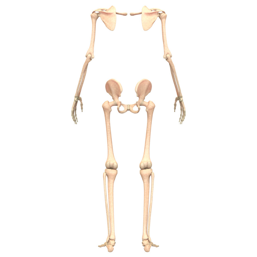
Appendicular skeleton
- Includes the bones of the upper and lower extremities, as well as the shoulder and pelvic girdles.
- Contains 126 bones in total.
- Responsible for movement and interaction with the environment.
Upper & Lower Extremities

Upper extremity
- Functions in grasping, manipulating objects, and fine motor control.
- Extends from the clavicle to the fingers, including the shoulder girdle (scapula and clavicle) and the upper limb (humerus, radius, ulna, carpals, metacarpals, phalanges).

Lower extremity
- Designed for weight-bearing, stability, and locomotion.
- Extends from the pelvic girdle to the toes, including the femur, patella, tibia, fibula, tarsals, metatarsals, and phalanges.