Day 1 – Trapezius, Levator scapula
Trapezius Muscle
The trapezius muscle is a large muscle of the back. It is a thin, flat muscle, and is the most superficial muscle of the posterior thoracic and cervical regions. The trapezius muscle got its name due to its shape. The left and right trapezius muscles are each triangular shaped, but together resemble a trapezoid (4-sided shape). This muscle has a broad origin which ranges from the external protuberance of the occipital bone (skull) down to the lower thoracic vertebrae. It is divided into upper, middle and lower sections.
- Upper trapezius (also called descending part), elevates and upwardly rotates the scapula; elevates the clavicle; extends the neck; laterally flexes the upper C-spine.
- Middle trapezius (also called transverse part), retracts or adducts the scapula (pulls toward the midline).
- Lower trapezius (also called ascending part), depresses and upwardly rotates the scapula.
Because of the broad origin of the trapezius, each section has fibers that run in different directions and therefore create different actions on the scapula. All sections contribute to scapular stabilization. The trapezius creates movement at several joints in the shoulder and cervical regions, but primarily affects the scapulothoracic (ST) joint. The scapulothoracic joint is not a true anatomical joint, but rather a gliding joint formed between the concave surface of the anterior scapula and the convex surface of the posterior thoracic cage. The trapezius is also a postural muscle. Poor posture such as forward head posture can result in increased muscle tension in the trapezius, especially the upper trapezius.
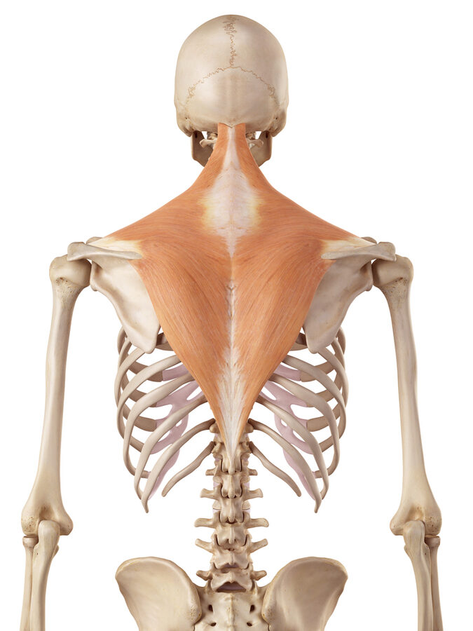

Trapezius
Origin: External occipital protuberance, medial third of superior nuchal line, ligamentum nuchae, spinous processes of vertebrae C7-T12
Insertion: Lateral third of clavicle, acromion process and upper crest of the scapular spine
Action: Elevation, depression, retraction or upward rotation of scapula (depending on which part of the muscle contracts). Cervical extension (upper trapezius).
Innervation: Accessory nerve (CNXI), and cervical nerves (C3,4)
Levator Scapula Muscle
The levator scapulae muscles are superficial muscles of the upper thoracic and posterior cervical regions. The superior end of the levator scapula is covered by the sternocleidomastoid (SCM) muscle. And at its inferior end, the levator scapula lies deep to the trapezius muscle. The middle part of the levator scapula is the easiest to palpate, and is located in the posterior triangle of the neck. The levator scapula is named according to its primary function which is to elevate the scapula (shoulder blade). It also rotates the scapula downward which means that the glenoid fossa at its lateral aspect tilts downward. If the shoulder blade is stabilized by the surrounding muscles, the levator scapula can function to laterally flex and extend the neck.
There are several conditions associated with dysfunction of the levator scapula muscle. This muscle is also susceptible to developing trigger points and muscle hypertension, especially in people with chronic stress or anxiety. Poor posture such as forward head posture and posture abnormalities such as scoliosis and torticollis can result in levator scapula dysfunction. Poor ergonomics can also lead to muscle tightness and dysfunction of the levator scapula. Massage therapists can use assisted stretching techniques in addition to massage to help lengthen, relax, and restore the normal function of this muscle.
- Cervical myofascial pain
- Cervicogenic headaches
- Trigger points (especially at the muscle’s attachment points)
- Fibromyalgia
- Levator scapula syndrome


Levator Scapula
Origin: Posterior tubercles of the transverse processes of C1-4 vertebrae
Insertion: Medial (vertebral) border of scapula, between the superior angle and root of the scapular spine
Action: Elevation and downward rotation of scapula
Innervation: Dorsal scapular nerve (C5), and cervical nerves (C3,4)
Learning in Action
- Demonstrate the actions of each muscle.
- Locate the origin and insertion of the trapezius and levator scapula muscles on yourself or a study partner.
- Palpate each muscle from origin to insertion (as much as possible). As you palpate, alternately perform a gentle contraction and relaxation of the muscle to help you differentiate this muscle from the surrounding tissues.
- Find the following bony landmarks and say the name of structures (out loud) as you palpate them. Also say how each feature relates to today’s two muscles. For example, “This is the acromion process and is an insertion point…“.
- External occipital protuberance of the skull
- Spinous processes of the C7-T12 vertebrae
- Transverse processes of C1-4 vertebrae
- Spine of the scapula
- Medial border and superior angle of the scapula
- Clavicle
- Acromion process of the scapula
- Draw the muscles.
- Study the trapezius muscle image for ~1 minute.
- Sketch the trapezius and surrounding structures on a sheet of paper, using the image as a reference. Make this sketch as accurate as possible but complete this in 1 minute or less. Compare your sketch to your reference image.
- Now turn your paper over and sketch the muscle without looking at any other images. Include as much detail as you can remember, but complete this in 1 minute or less. Compare your sketch to your reference image. Note any errors in your mental image of the muscle. Especially where the muscle attaches to bone, and the size and position relative to other structures around it.
- Go back and label the origin, insertion, and any key features of this muscle or surrounding structures. Add more detail if you need to.
- Repeat this process for the levator scapula muscle.
- Create your muscle flashcards for the trapezius and levator scapula muscles.
How muscles are named
There are 8 main ways that skeletal muscles are named. The names of many muscles use a combination of these naming strategies. Here are the 8 ways and some examples:
- Shape: muscles named for their shape include the trapezius and deltoid.
- Action: levator scapula, adductor longus, extensor digitorum, and flexor hallucis longus.
- Attachment points: examples include the coracobrachialis, and the sternocleidomastoid (SCM) muscles.
- Fiber direction: the rectus abdominis and rectus femoris muscles (“rectus” means straight or parallel).
- Relative size: gluteus maximus, gluteus medius and glutes minimus. Pectoralis major and minor. Rhomboid major and minor.
- Relative position: vastus lateralis and vastus medialis.
- Other physical characteristics: 2 heads or origins (biceps brachii or biceps femoris). Or 3 heads, triceps brachii.
- Location: in a certain region, like the upper arm or brachial region (brachialis).